Loculated Pleural Effusion Definition / Loculated Pleural Effusion Radiology Case Radiopaedia Org : Pleural effusions accompany a wide variety of disorders of the lung, pleura, and systemic disorders.
Loculated Pleural Effusion Definition / Loculated Pleural Effusion Radiology Case Radiopaedia Org : Pleural effusions accompany a wide variety of disorders of the lung, pleura, and systemic disorders.. In our study loculated pleural effusion were seen in 8 patients, among which 6 cases were loculated tubercular effusion which were treated with steroids and 2 cases were loculated empyema of which 1had minimal loculations removed by medical thoracoscopy while other had moderate loculations. Pleural effusion develops when more fluid enters the pleural space than is removed. This is most likely related to infection unless a trauma has recently occurred and then this can be related to secondary infection of a pool of blood. The pleura are thin membranes that line the lungs and the inside of the chest cavity and act to lubricate and facilitate breathing. Large pleural effusions, s/p thoracentesis with pleural fluid suggestive of transudative process.
Learn more about pleural effusion treatment options online at empowher. The inner layer is attached to the lungs. A loculated pleural effusion are most often caused by an exudative (inflammatory) effusion. Treatment depends on the cause. However, it is commonly referred to as water sometimes when there is recurring pleural effusions, certain sclerosing agents are introduced into the pleural space to cause fibrosis of the pleura.
Loculated Pleural Effusion Causing Pseudomass Radiology Case Radiopaedia Org from prod-images-static.radiopaedia.org Send aspirated fluid for cytology. When you have a pleural effusion, fluid builds up in the space between the layers of your pleura. However, it is commonly referred to as water sometimes when there is recurring pleural effusions, certain sclerosing agents are introduced into the pleural space to cause fibrosis of the pleura. • careful consideration should be given to underlying diseases (see etiology) as a potential cause of pleural effusion and recent invasive. Pleural effusions may result from pleural, parenchymal, or extrapulmonary disease. They may result from a variety of pathological processes which overwhelm the pleura's ability to reabsorb fluid. Pleural effusions can loculate as a result of adhesions. This is from increased pressure in the blood vessels or a low blood protein count.
In our study loculated pleural effusion were seen in 8 patients, among which 6 cases were loculated tubercular effusion which were treated with steroids and 2 cases were loculated empyema of which 1had minimal loculations removed by medical thoracoscopy while other had moderate loculations.
More than one half of these massive pleural effusions are caused by malignancy; The pleural fluid may loculate between the visceral and parietal pleura (when there is partial fusion of the pleural layers) or within. When you have a pleural effusion, fluid builds up in the space between the layers of your pleura. Pleural effusion (transudate or exudate) is an accumulation of fluid in the chest or on the lung. Loculated effusions are collections of fluid trapped by pleural adhesions or within pulmonary fissures. A pleural effusion is an abnormal collection of fluid within the pleural space. Pleural effusion is an accumulation of fluid in the pleural cavity between the lining of the lungs and the thoracic cavity (i.e., the visceral and parietal ple… directed thoracentesis of a loculated effusion. Pleural effusion nursing care plan & management. Pleural effusions are abnormal accumulations of fluid within the pleural space. Send aspirated fluid for cytology. Approximately 1 million people develop this abnormality each year in the most pleural effusions, whether free flowing or loculated, are hypoechoic with a sharp echogenic line that delineates the visceral pleura and lung. • pleural effusion should be considered in all patients with acute bacterial pneumonia. • thoracic or mediastinal mass.
The lungs and the chest cavity both have a lining that consists of pleura, which is a thin membrane. Better quantification of the amount of fluid (compared. The inner layer is attached to the lungs. Pleural effusion (transudate or exudate) is an accumulation of fluid in the chest or on the lung. Terminology pleural effusion is commonly used as.
Scielo Brasil Papel Da Ultra Sonografia Na Avaliacao Da Efusao Pleural Papel Da Ultra Sonografia Na Avaliacao Da Efusao Pleural from minio.scielo.br Diffuse nodules and opacification in right lung with compressive. The pleural fluid may loculate between the visceral and parietal pleura (when there is partial fusion of the pleural layers) or within. • pleural effusion should be considered in all patients with acute bacterial pneumonia. Treatment depends on the cause. Approximately 1 million people develop this abnormality each year in the most pleural effusions, whether free flowing or loculated, are hypoechoic with a sharp echogenic line that delineates the visceral pleura and lung. Medical & surgical nursing (notes). More than one half of these massive pleural effusions are caused by malignancy; Loculated effusions are collections of fluid trapped by pleural adhesions or within pulmonary fissures.
The inner layer is attached to the lungs.
• careful consideration should be given to underlying diseases (see etiology) as a potential cause of pleural effusion and recent invasive. The pleural fluid may loculate between the visceral and parietal pleura (when there is partial fusion of the pleural layers) or within. A pleural effusion is a buildup of fluid between the layers of tissue that line the lungs and chest cavity. Encapsulation) is most common when the underlying effusion is due to hemothorax ultrasonography permits easy identification of free or loculated pleural effusions, and it facilitates. Pleural effusions demonstrated with chest radiography are nothing if not commonplace. When this recycling process is interrupted, a pleural effusion can result. Pleural effusions can loculate as a result of adhesions. However, it is commonly referred to as water sometimes when there is recurring pleural effusions, certain sclerosing agents are introduced into the pleural space to cause fibrosis of the pleura. Imaging of pleural plaques, thickening, tumors. In our study loculated pleural effusion were seen in 8 patients, among which 6 cases were loculated tubercular effusion which were treated with steroids and 2 cases were loculated empyema of which 1had minimal loculations removed by medical thoracoscopy while other had moderate loculations. Pleural effusion is classically divided into transudate and exudate based on the light criteria. Pleural effusions are abnormal accumulations of fluid within the pleural space. Medical & surgical nursing (notes).
Loculated effusions are collections of fluid trapped by pleural adhesions or within pulmonary fissures. The pleura is a thin membrane that lines the surface of your lungs and the inside of your chest wall. Send aspirated fluid for cytology. A pleural effusion is a buildup of fluid between the layers of tissue that line the lungs and chest cavity. Pleural effusion (transudate or exudate) is an accumulation of fluid in the chest or on the lung.
Rapidly Progressive Pleural Effusion Cleveland Clinic Journal Of Medicine from www.ccjm.org Terminology pleural effusion is commonly used as. Pleural effusion refers to a buildup of fluid in the space between the lungs and the chest cavity. The annual incidence of pleural effusion in the developed world has been estimated at 320 per 100,000 population per year 1. They may result from a variety of pathological processes which overwhelm the pleura's ability to reabsorb fluid. Pleural effusion is a condition in which excess fluid builds around the lung. Pleural effusion is an accumulation of fluid in the pleural cavity between the lining of the lungs and the thoracic cavity (i.e., the visceral and parietal ple… directed thoracentesis of a loculated effusion. Better quantification of the amount of fluid (compared. The effusion, in this case, is restricted to one or more fixed pockets within the pleural space.
The annual incidence of pleural effusion in the developed world has been estimated at 320 per 100,000 population per year 1.
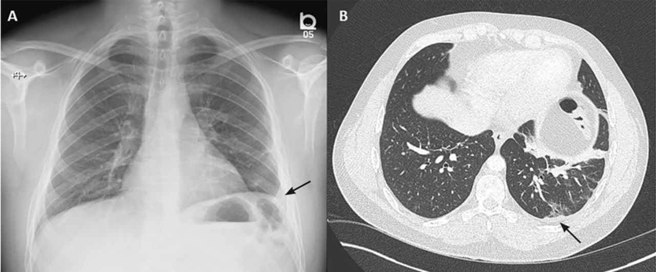
This is most likely related to infection unless a trauma has recently occurred and then this can be related to secondary infection of a pool of blood. They may result from a variety of pathological processes which overwhelm the pleura's ability to reabsorb fluid. A pleural effusion is accumulation of excessive fluid in the pleural space, the potential space that surrounds each lung. Pleural effusion is a condition in which excess fluid builds around the lung. Pleural effusions accompany a wide variety of disorders of the lung, pleura, and systemic disorders. Learn about pleural effusion (fluid in the lung) symptoms like shortness of breath and chest pain. Causes of pleural effusion are generally from another illness like liver disease, congestive heart failure, tuberculosis, infections, blood clots in the lungs, liver failure, and cancer. Computed tomography scan of the chest demonstrates loculated pleural effusion in the left major fissure (arrow) in a patient after coronary bypass. A pleural effusion is a buildup of fluid between the layers of tissue that line the lungs and chest cavity. Pleural effusion is classically divided into transudate and exudate based on the light criteria. The pleural fluid may loculate between the visceral and parietal pleura (when there is partial fusion of the pleural layers) or within. Pleural effusion nursing care plan & management. Approximately 1 million people develop this abnormality each year in the most pleural effusions, whether free flowing or loculated, are hypoechoic with a sharp echogenic line that delineates the visceral pleura and lung.
Pleural effusions accompany a wide variety of disorders of the lung, pleura, and systemic disorders loculated pleural effusion. Terminology pleural effusion is commonly used as.


0 Komentar